

Your Browsing History

Meet Your Seller

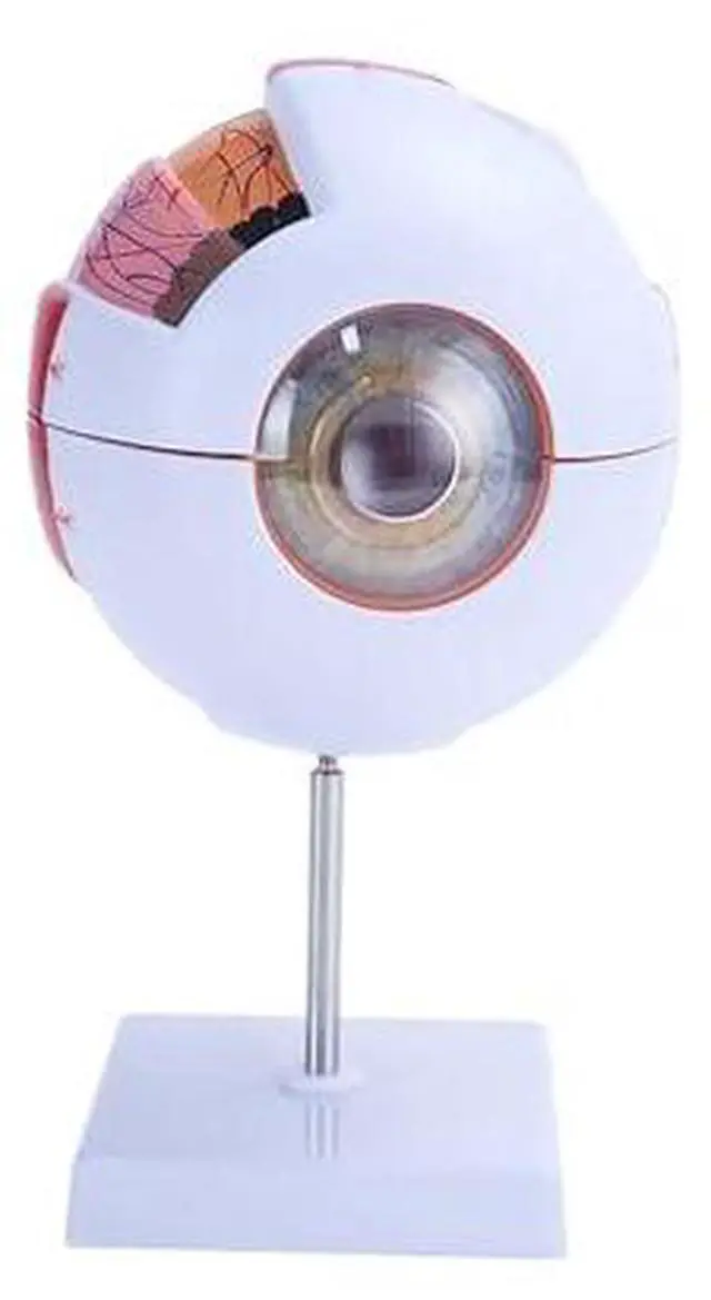
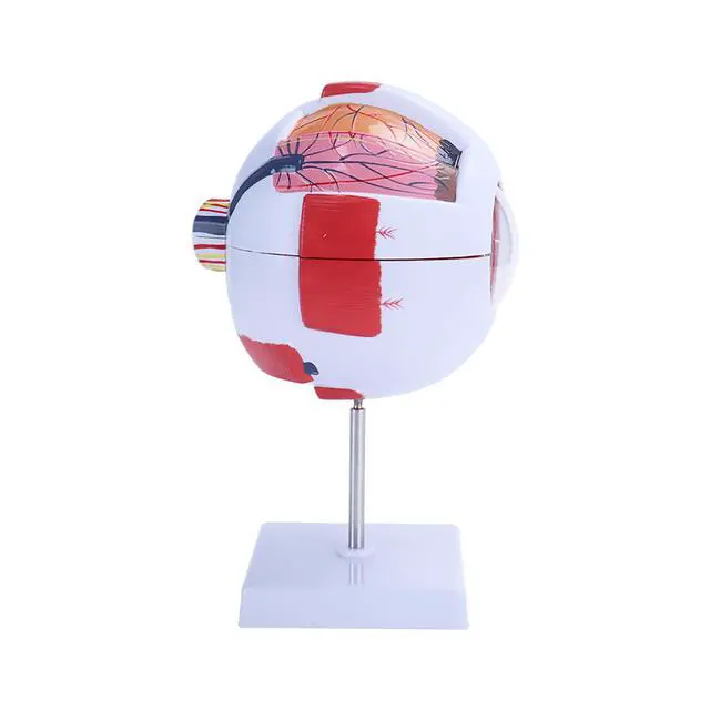
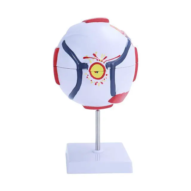
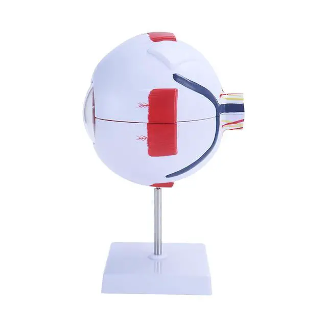
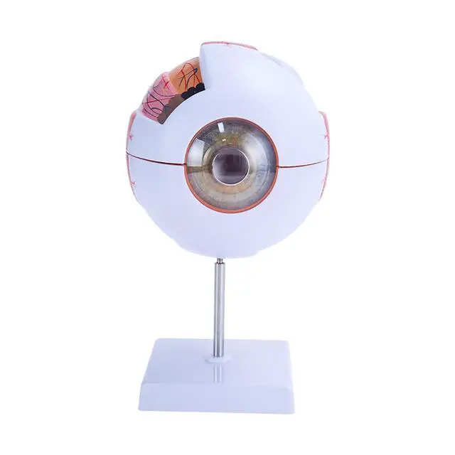


AMUREKE 6X Human Eye Model Enlarged Anatomical Eyeball Model for Science Classroom Study Display Medical Education with Removable Stand
- [Detailed Anatomy at 6x Magnification]The model features 6x magnification to clearly show the intricate details of the human eye. Designed for comprehensive learning, it is an invaluable tool for student education and anatomical research.
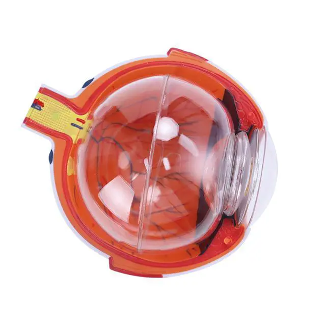
- [PROFESSIONALIZATION]This model is used to demonstrate the anatomical structure of the human eye, such as the three membranes of the eyeball wall (outer, middle and inner membranes) as well as the main refractive bodies, lens and vitreous body that fill the interior. This eyeball model is suitable for use as a visual aid in teaching.
- [Anatomically accurate]The anatomical model of the eyeball contains seven parts: upper and lower part of the eye, cornea, iris, lens, vitreous body and stent. It accurately represents the anatomy of the human eye, emphasizing key features such as the three layers of the eye wall and the main refractive components.
- [Widely Used] The model can be used for science education, student learning, demonstration and medical teaching. It is suitable for professionals such as physiotherapists, radiographers and practicing physicians. Its adaptability makes it suitable for a variety of educational and medical settin
- [Practical Design with Removable Parts] Equipped with a sturdy base, the model is easy to install and remove for demonstration purposes. Its detachable design allows for closer examination of the internal anatomy of the eye, enhancing the learning experience.
+
+
Overview
Specs
Reviews

Any questions? Our AI mode will help you find out quickly.
Try me now
Product Features
High detail display: with 6 times magnification function, it can clearly present the fine structure of the human eye and help comprehensive learning, which is a powerful tool for students' education and anatomical research.
Professional Teaching Aid: It is used to show the anatomical structure of the human eye, such as the three membranes of the eyeball wall (outer, middle and inner membranes) and the main refractive bodies, lens and vitreous body in the eye, etc. It is suitable for teaching visualisation aid.
Precise anatomical reproduction: The anatomical model of the eyeball contains seven parts: the upper and lower part of the eye, cornea, iris, lens, vitreous and stent, which accurately presents the anatomical structure of the human eye and highlights the key features of the three membranes of the wall of the eyeball as well as the main refractive components.
Wide range of application scenarios: suitable for science education, student learning, demonstration and medical teaching, and can also be used by professionals such as physiotherapists, radiologists and medical practitioners, suitable for a variety of educational and medical scenarios.
Practical and detachable design: Equipped with a stable base, easy to install and dismantle the display. The detachable design is convenient for in-depth observation of the internal anatomy of the eye, enhancing the learning experience.
Product Parameters
Magnification: 6x
Components: upper and lower part of the eye, cornea, iris, lens, vitreous, support
Applicable scenes: science education, student learning, demonstration, medical teaching
Product information
| Package Dimensions | 7 x 5 x 4 inches; 1 Pounds |
| Date First Available | April 29, 2025 |
| Manufacturer | Generic |
Warranty & Returns
Warranty, Returns, And Additional Information
Warranty
- Please contact the Seller directly for warranty information. Warranty information may also be found on the Manufacturer's website.
- CONTACT
Return Policies
- Return for refund within: 30 days
- Return for replacement within: non-replaceable
- This item is covered by UGETSEE LIMITED Return Policy
Manufacturer Contact Info
- For a directory of all our manufacturers, please click below.
- Manufacturers Directory
LOADING...